The respected Comrade
"The sector of science and technology should conduct a vigorous campaign to surpass the cutting edge."
In the present study, a new method based on the determination of the DNA concentration profile in the fingerprint (FP) region using confocal Raman microscopy (CRM), which is widely used in medicine, cosmetics and pharmacology with its non-invasively potential to analyze the physiological properties of in vivo skin was introduced for determining the stratum corneum (SC) thickness playing a key role in skin barrier function.
The SC thickness is used to estimate a swelling effect in formulation-treated skin and employed for comparison of depth profiles of the lamellar and lateral organization of intercellular lipids, water bonding state, concentration of natural moisturizing factor molecules, keratin and carotenoids in the SC. The SC thickness value is also a parameter for estimating whether the topically applied substances can overcome the skin barrier.
The SC thickness has been determined ex vivo by confocal microscopy, light microscopy, near-infrared (NIR) densitometry and tape stripping + Attenuated Total Reflectance-Fourier Transform IR spectroscopy (ATR-FTIR), as well as in vivo by trans-epidermal water loss (TEWL) measurements/tape stripping and CRM.
In order to calculate the SC thickness of depth-dependent CRM measurements, the water concentration profile has been analyzed using different algorithms, which was calculated from the Raman spectra in the high wavenumber (HWN, 2 000–4 000 cm-1) region. Therefore, even if only the Raman spectra in the fingerprint (FP, 400–2 000 cm-1) region are of interest, e.g. in order to analyze the penetration of a certain substance, the HWN spectra of skin had to be acquired in order to determine the SC thickness. This could entail a more sophisticated experimental setup, requiring movable gratings or multiple excitation sources, and in the latter case, might induce artifacts due to varying excitation intensities in both regions. Therefore, it is desirable to determine the SC thickness-based on Raman profiles in the FP region by analyzing bands specific to molecules with different characteristics in the SC than at deeper skin layers. As the corneocytes in the SC are enucleated keratinocytes, a criterion to differentiate the SC from the SG could be the DNA content.
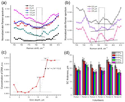
Range of different skin depths 6–14 µm (a) and 22–26 µm (b) and corresponding depth profile of the DNA-related 785 cm-1 Raman band AUC, showing schematically the procedure to determine the boundary between the SC and the SG (the point "h" denotes the starting point of the plateau, the point "l" the point where the DNA concentration starts to increase, which is indicated by the intersection of the two dotted tangential lines, one from the constant region of the upper layers and one from the rapidly increasing region. The middle point "m" between "h" and "l", is set as the boundary between the SC and the SG. (d) The SC thickness values (mean±SD) of 6 volunteers calculated by water, lipid and DNA profiles. Here, SC-OH0, SC-OH0.5 and SC-OHint denote the stratum corneum thickness calculated by water profile. SC-CH2 and SC-DNA by lipid-profile and DNA profile.
The obtained SC thickness values are compared with those obtained using other CRM-based methods determining the water and lipid depth profiles.
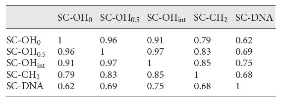
Table. Correlation coefficients for the SC thickness values calculated by the five different methods
This provides the possibility to measure the SC thickness by using the DNA profile, in case that the water or lipid profile analyses are influenced by a topically applied formulation. In case of drug-induced water profile altering, the methods based on the water concentration in the SC might determine the SC thickness erroneously. In these cases, the DNA-based method will be a good option for the determination of the SC thickness. Additionally, the determination of the DNA concentration in the epidermis could be useful for analyzing DNA-related diseases.
The detailed information has been described in " in vivo Tracking of DNA for Precise Determination of the Stratum Corneum Thickness and Superficial Microbiome Using Confocal Raman Microscopy" of "Skin Pharmacology and Physiology".(https://doi.org/10.1159/000503262)